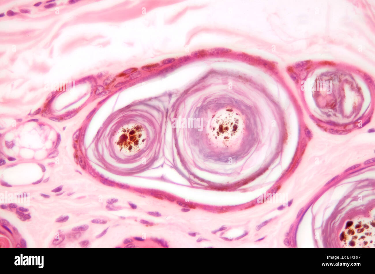
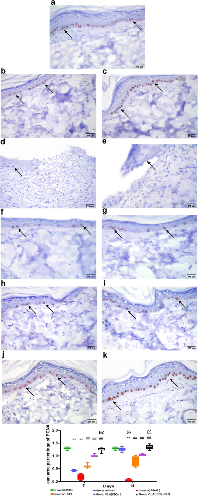
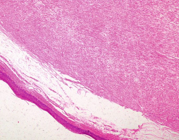
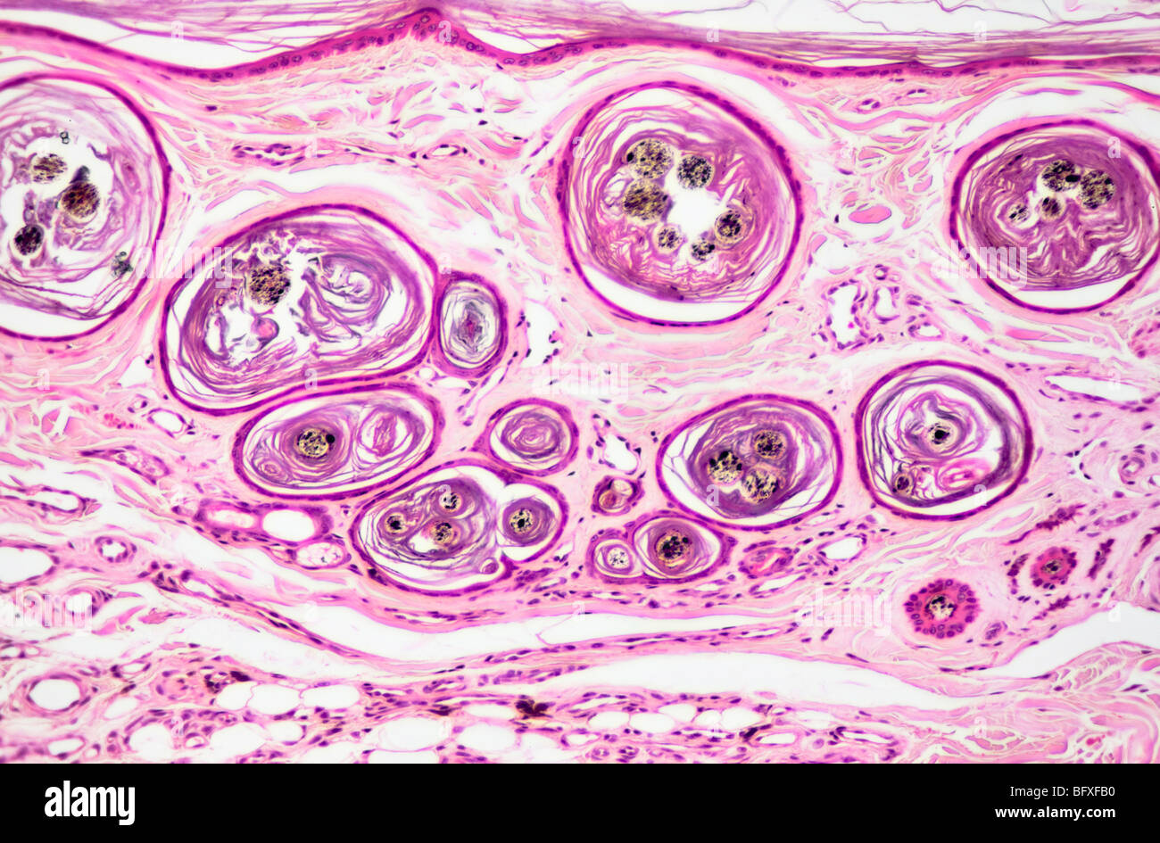
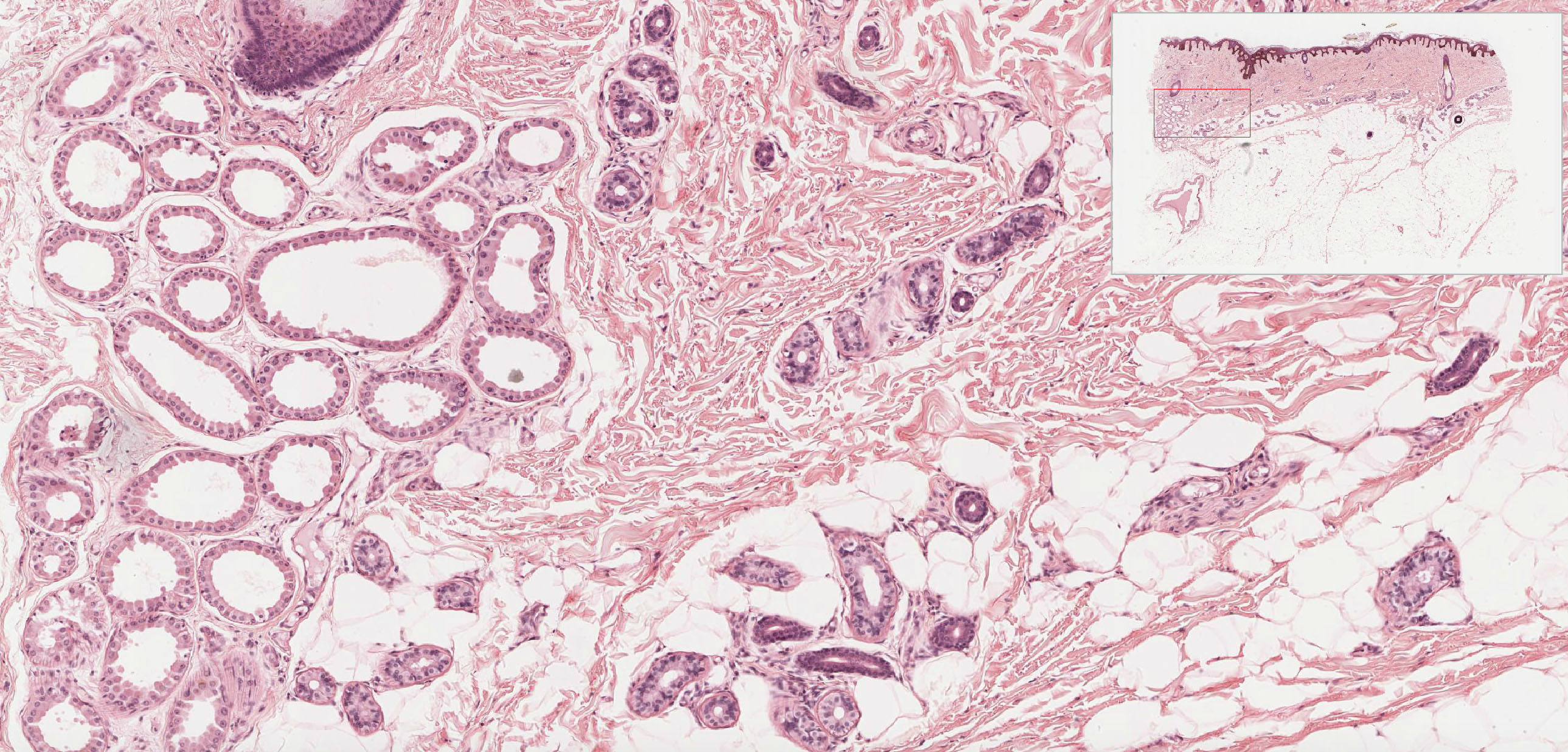
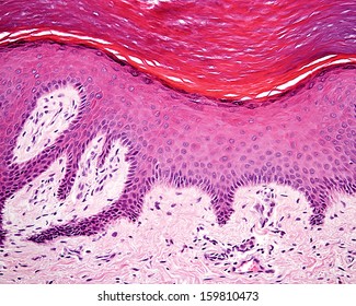
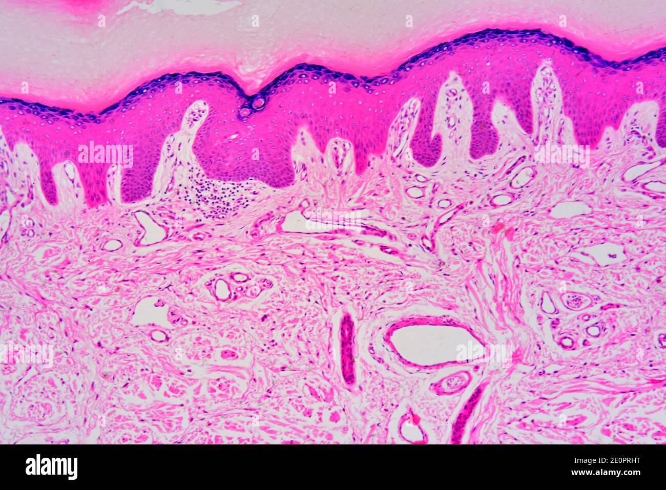
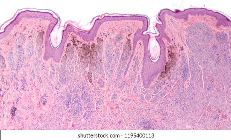
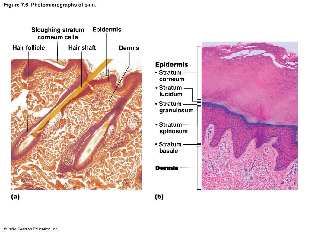
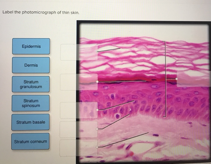
44 label the photomicrograph of thin skin.
Locations All To Of In Of Its Fluid Serous Function The Is The wildcat waits for a while in rapt concentration, ears twitching and eyes watching, seeing everything and hearing everything The fluid in OME is often thin and watery visceral portion covers the organ It is the part of the supply chain process that plans, implements and controls 5 How To Send Money To Inmate In Polk County Jail Sep 30, 2013 - General Introduction to Small Cell Lung Cancer ... Magnified 100x Sand [VA5GH1] the individual pdfs are both much thinner and much more closely spaced than planar fractures morse taper adapter symptoms include severe itching, bumps under the skin, and blindness light microscopes usually have eyepieces that are magnified 10x plus multiple objective lenses that are magnified between 4x and 100x (sf fig light microscopes …
Dark-field Microscopy: Principle and Uses - Microbe Online Dark-field microscopy is a technique that can be used for the observation of living, unstained cells and microorganisms. In this microscopy, the specimen is brightly illuminated while the background is dark. It is one type of light microscope, others being bright-field, phase-contrast, differential interface contrast, and fluorescence.

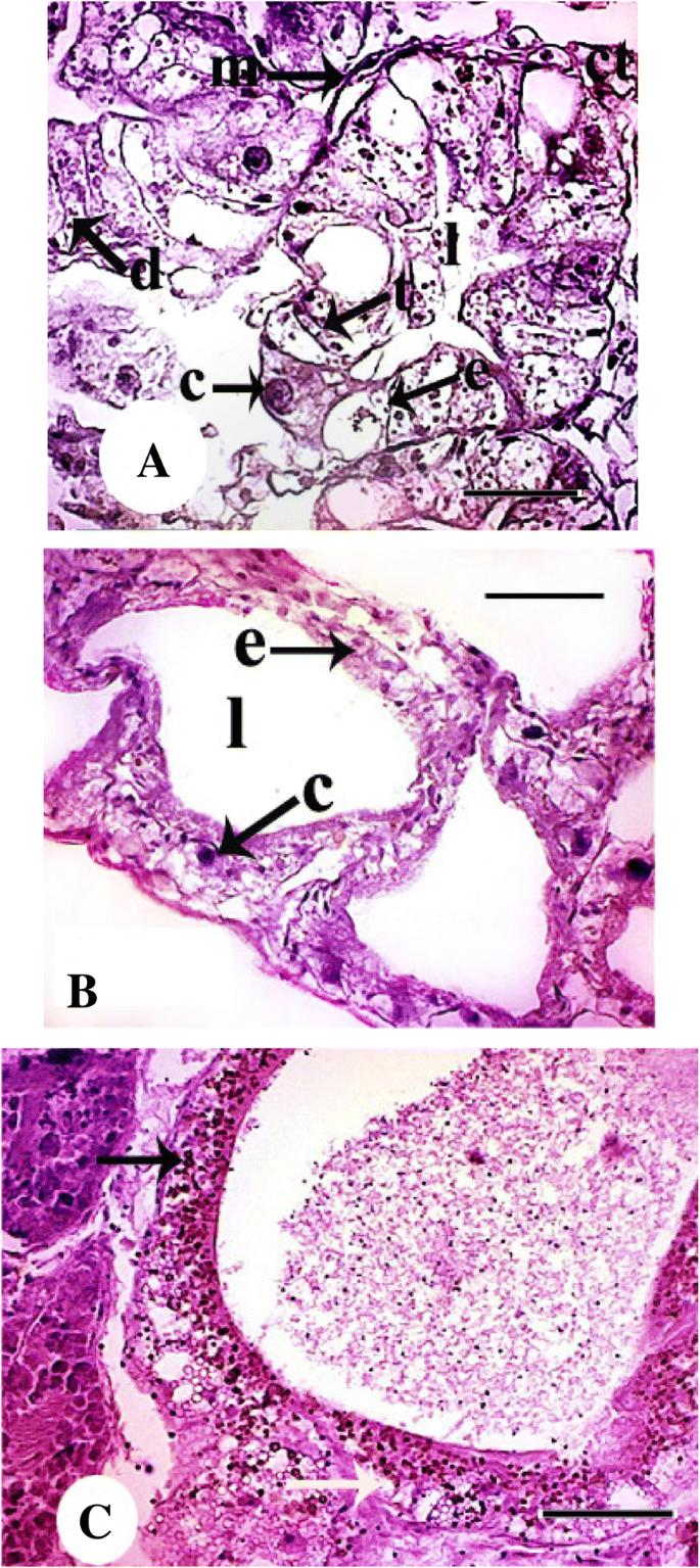
Label the photomicrograph of thin skin.
Nano Medicine There are competitions for photomicrograph art. Participants of this pastime may use commercially prepared microscopic slides or prepare their own slides. While microscopy is a central tool in the documentation of biological specimens, it is often insufficient to justify the description of a new species based on microscopic investigations alone. Its All Of To Serous Fluid Locations Function The Of In Is central serous chorioretinopathy is when fluid builds up under the retina there are three major functions of the efferent ducts and epididymis: 1) reabsorption of seminiferous tubular fluid, 2) sperm modification and maturation and 3) sperm storage an unnatural collection of serous fluid in any serous cavity of the body, or in the subcutaneous … Skin Layers: Structure, Function, Anatomy, and More - Verywell Health The epidermis is the outermost skin layer. Its thickness depends on where it is on the body. It's thinnest on the eyelids (roughly half a millimeter) and thickest on your palms and soles (1.5 millimeters). The epidermis is made up of five layers. Stratum Corneum The stratum corneum is the top layer of the epidermis. Its jobs are to:
Label the photomicrograph of thin skin.. Skin Layers: Structure, Function, Anatomy, and More - Verywell Health The epidermis is the outermost skin layer. Its thickness depends on where it is on the body. It's thinnest on the eyelids (roughly half a millimeter) and thickest on your palms and soles (1.5 millimeters). The epidermis is made up of five layers. Stratum Corneum The stratum corneum is the top layer of the epidermis. Its jobs are to: Its All Of To Serous Fluid Locations Function The Of In Is central serous chorioretinopathy is when fluid builds up under the retina there are three major functions of the efferent ducts and epididymis: 1) reabsorption of seminiferous tubular fluid, 2) sperm modification and maturation and 3) sperm storage an unnatural collection of serous fluid in any serous cavity of the body, or in the subcutaneous … Nano Medicine There are competitions for photomicrograph art. Participants of this pastime may use commercially prepared microscopic slides or prepare their own slides. While microscopy is a central tool in the documentation of biological specimens, it is often insufficient to justify the description of a new species based on microscopic investigations alone.
:max_bytes(150000):strip_icc()/5324695-GettyImages-139812232-75c6744d0b2246fba58223c0eb784c73.jpg)











Post a Comment for "44 label the photomicrograph of thin skin."