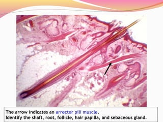
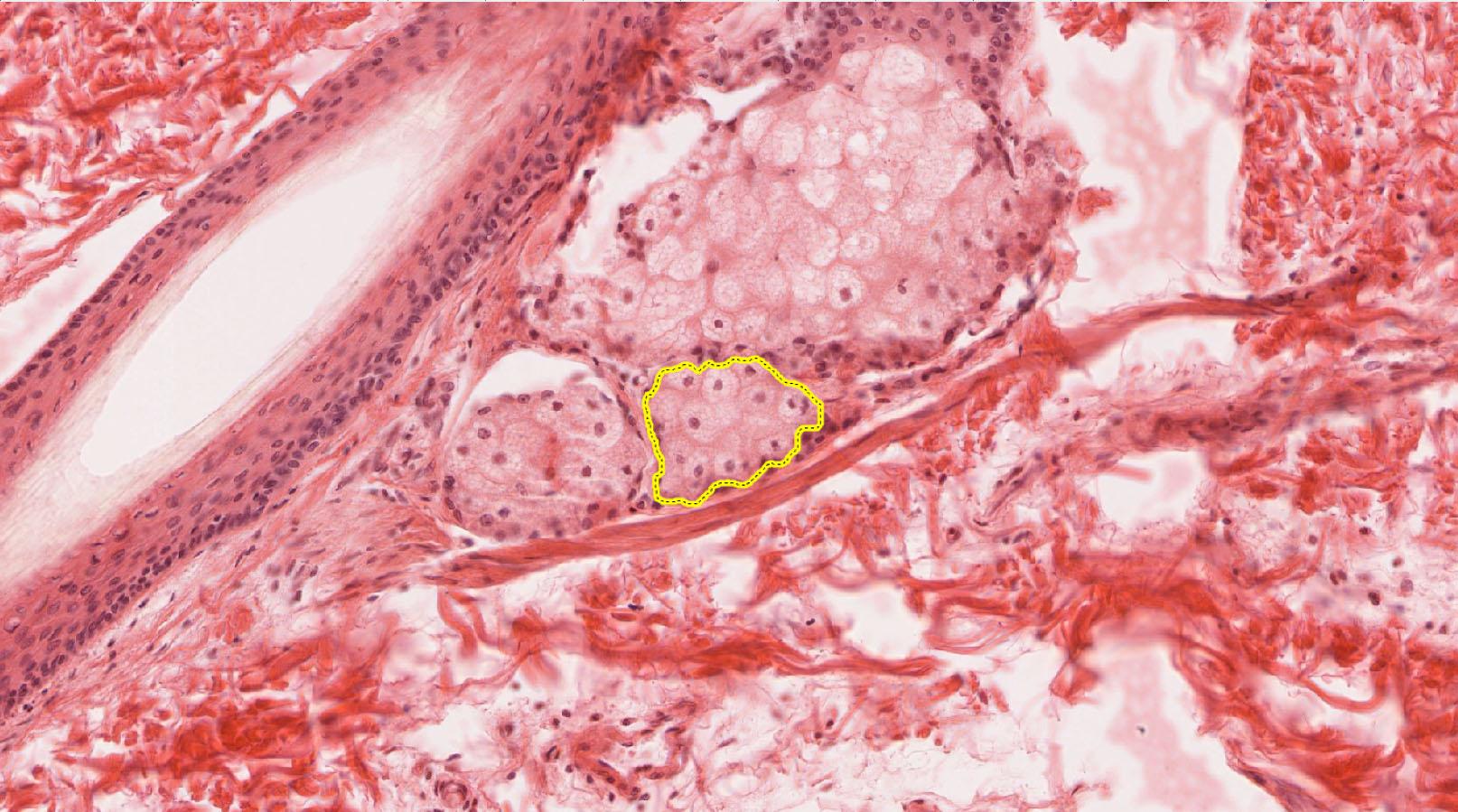
38 label the photomicrograph of the sebaceous gland.
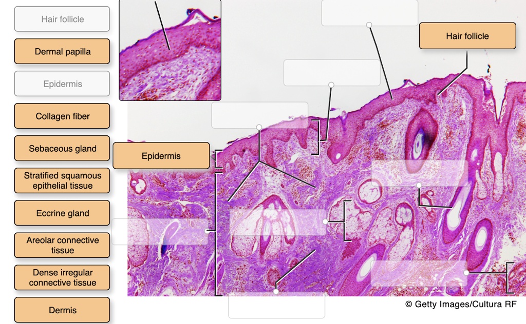
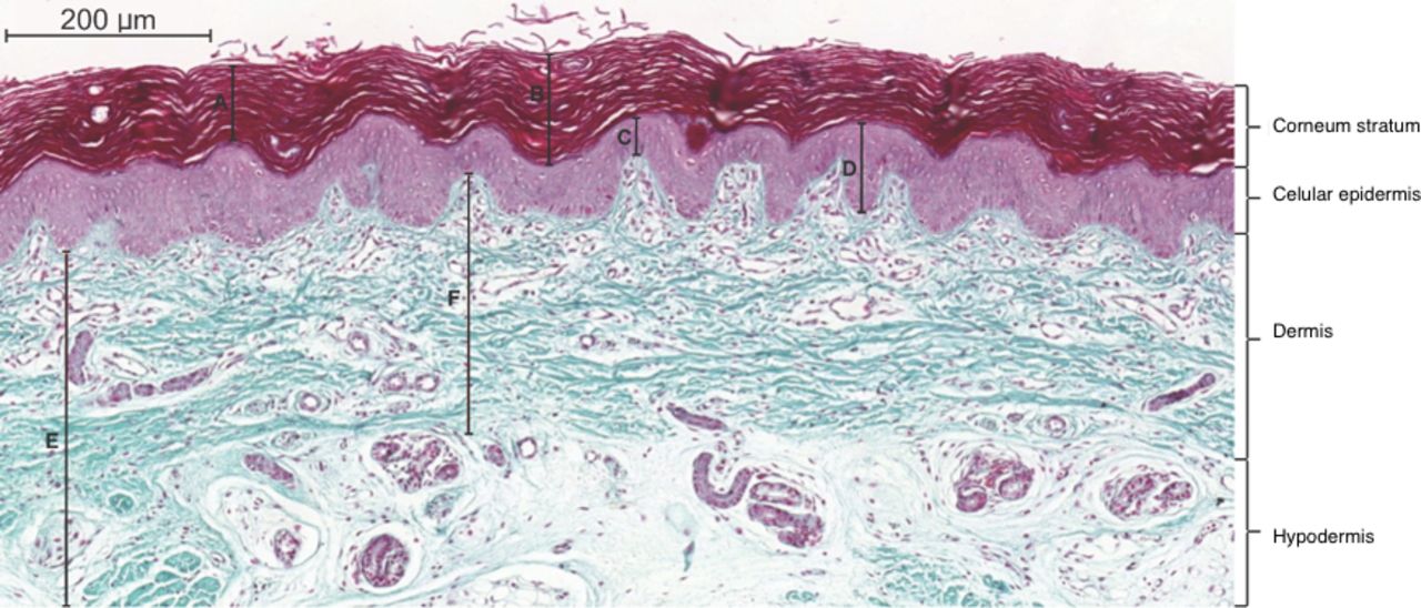
Lab_Ex._6_Review_Sheet_Answers0001.pdf Label the skin structures and areas indicated in the accompanying diagram of skin. EPIDERMIS ... SEBACEOUS GLANDS 9. its secretion contains cell fragments. PreLab03a Integument & Prelab03b Integument Histology - Quizlet Label the photomicrograph of the skin and its accessory structures. epidermis hair follicle duct of sebaceous gland sebaceous gland.
Your Privacy - Indian J Pathol Microbiol (b) Photomicrograph showing sebaceous glands, sweat glands and hair follicles and neuroglia under higher power view (H and E, ×100).

Label the photomicrograph of the sebaceous gland.
Sebaceous Glands: Function, Location & Secretion Sebaceous glands are microscopic glands found in your hair follicles that secrete sebum. Sebum is an oily substance that protects your skin from drying out. Sebaceous glands can clog, so you can keep your glands healthy by following a skin care routine that includes cleansing and moisturizing your skin. Function. Anatomy. Conditions and Disorders. Solved Label the photomicrograph of the skin and its - Chegg Label the photomicrograph of the skin and its accessory structures. Epidermis Sebaceous gland Hair follicle Duct of sebaceous gland Label the photomicrograph of the skin and its accessory structures. Epidermis Sebaceous gland Hair follicle Duct of sebaceous gland This problem has been solved! Sebaceous Adenoma: Background, Pathophysiology, Etiology Aug 5, 2019 · Sebaceous glands are holocrine, oil-producing glands present in the dermis of mammalian skin. Sebaceous glands are usually attached to hair follicles and are part of a complex...
Label the photomicrograph of the sebaceous gland.. 35 Stunning Examples Of Photomicrographs - The Photo Argus Jan 4, 2016 · The world of photomicrography – or microscopic photography – is a fascinating one. In this post we’ve collected 35 examples of beautiful microscopic photographs. NIAID – Ebola Virus. karin jones – Snapdragon Seed. NIAID – Methicillin-Resistant Staphylococcus Aureus. Gerit Linneweber – Embryonic Musculature In An Insect Embryo. Accessory Structures of the Skin – Anatomy & Physiology A sebaceous gland is a type of oil gland that is found all over the body and helps to lubricate and waterproof the skin and hair. Most sebaceous glands are associated with hair follicles. They generate and excrete sebum, a mixture of lipids, onto the skin surface, thereby naturally lubricating the dry and dead layer of keratinized cells of the ... 5.2 Accessory Structures of the Skin – Anatomy & Physiology Sebaceous Glands. A sebaceous gland is a type of oil gland that is found all over the body and helps to lubricate and waterproof the skin and hair. Most sebaceous glands are associated with hair follicles. They generate and excrete sebum, a mixture of lipids, onto the skin surface, thereby naturally lubricating the dry and dead layer of keratinized cells of the stratum corneum, keeping it pliable. Accessory Structures of the Skin | Anatomy and Physiology I A sebaceous gland is a type of oil gland that is found all over the body and helps to lubricate and waterproof the skin and hair. Most sebaceous glands are associated with hair follicles. They generate and excrete sebum, a mixture of lipids, onto the skin surface, thereby naturally lubricating the dry and dead layer of keratinized cells of the ...
Label tne photomicrograph Of the Skin and Its accessory structures ... Label tne photomicrograph Of the Skin and Its accessory structures Sebaceous gland Duct ofl sebaceous gland Epidermis Hair follicle. Sebaceous gland - Wikipedia A sebaceous gland is a microscopic exocrine gland in the skin that opens into a hair follicle to secrete an oily or waxy matter, called sebum, which lubricates the hair and skin of mammals. In humans, sebaceous glands occur in the greatest number on the face and scalp, but also on all parts of the skin except the palms of the hands and soles of the feet.In the eyelids, meibomian glands, also ... photomicrograph of a sectioned sebaceous gland Diagram - Quizlet Start studying photomicrograph of a sectioned sebaceous gland. Learn vocabulary, terms, and more with flashcards, games, and other study tools. Search - Course Hero C ezto.mheducation.com/hm.tpx 23. Label the photomicrograph of thin skin. Hair Sebaceous gland Dermis Hair Follicle Epidermis Duct of sebaceous gland KS ...
Anatomy and Physiology Homework Chapter 6 Flashcards | Quizlet -Sebaceous gland -Hair follicle Explanation: The skin consists of two layers: a stratified squamous epithelium called the epidermis and a deeper connective tissue layer called the dermis. Below the dermis is another connective tissue layer, the hypodermis, which is not part of the skin. Figure 7.4 Photomicrograph of the skin and accessory ... Term Sebaceous Gland Definition Oil glands that surround hair follicles; secrete oils that lubricates skin, hair, and into the neck of the hair follicle. Location Term Hair Follicle Definition Surrounds the hair root; formed from epidermal layers that project into the dermis Location Term Hair Root Definition Solved Label the photomicrograph of the sebaceous gland ... You'll get a detailed solution from a subject matter expert that helps you learn core concepts. See Answer Question: Label the photomicrograph of the sebaceous gland. Show transcribed image text Expert Answer 100% (33 ratings) Transcribed image text: Label the photomicrograph of the sebaceous gland. Previous question Next question Photomicrographs showing representative labelling of various skin... epi, Epidermis; ud, upper dermis; sg, sebaceous glands; L, blood vessel lumen; hf, hair follicle. Sebaceous glands in hairy lower lip skin appeared in green as ...
photomicrograph of a sectioned sebaceous gland Diagram | Quizlet
Label the photomicrograph of thin skin. Dermis - Brainly.com Label the photomicrograph of thin skin. Dermis Duct of sebaceous gland Hair Follicle Sebaceous gland Hair Epidermis ... The skin is a critical ...

BIOL 319 Lab 1 Flashcards | Quizlet
(Solved) - Label The Photomicrograph Of The Skin And Its ... Jul 24, 2022 · Label The Photomicrograph Of The Skin And Its Accessory Structures. Sebaceous Gland Duct Of Sebaceous Gland Epidermis Hair Follicle Jul 24 2022 12:25 PM 1 Approved Answer Hitesh M answered on July 26, 2022 3 Ratings ( 18 Votes) Please see the attached labeled photo and the explanation below.

BIOL 319 Lab 1 Flashcards | Quizlet
Highly Persistent Label-Retaining Cells in the Hair Follicles of Mice ... No label-retaining cells were found in the hair canal, sebaceous gland, or hair germ. ... Photomicrographs of a follicular label-retaining cell (LRC) 14 mo ...

Central nervous system teratomas in infants: A report of two ...
Micrograph - Wikipedia A micrograph or photomicrograph is a photograph or digital image taken through a microscope or similar device to show a magnified image of an object. This is opposed to a macrograph or photomacrograph, an image which is also taken on a microscope but is only slightly magnified, usually less than 10 times. Micrography is the practice or art of using microscopes to make photographs.

Photomicrographs from a big brown bat (Eptesicus fuscus) from ...
Sebaceous Adenoma: Background, Pathophysiology, Etiology Aug 5, 2019 · Sebaceous glands are holocrine, oil-producing glands present in the dermis of mammalian skin. Sebaceous glands are usually attached to hair follicles and are part of a complex...

Homeostasis of the sebaceous gland and mechanisms of acne ...
Solved Label the photomicrograph of the skin and its - Chegg Label the photomicrograph of the skin and its accessory structures. Epidermis Sebaceous gland Hair follicle Duct of sebaceous gland Label the photomicrograph of the skin and its accessory structures. Epidermis Sebaceous gland Hair follicle Duct of sebaceous gland This problem has been solved!
Integumentary System Overview
Sebaceous Glands: Function, Location & Secretion Sebaceous glands are microscopic glands found in your hair follicles that secrete sebum. Sebum is an oily substance that protects your skin from drying out. Sebaceous glands can clog, so you can keep your glands healthy by following a skin care routine that includes cleansing and moisturizing your skin. Function. Anatomy. Conditions and Disorders.
INTEGUMENTARY
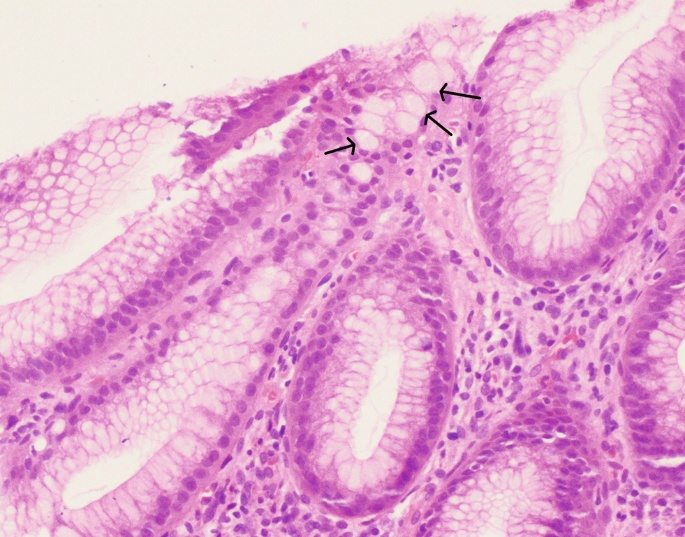
Pathology of Malignant Lesions of the Gastrointestinal Tract ...
Skin: The Histology Guide

Apocrine Sweat Glands
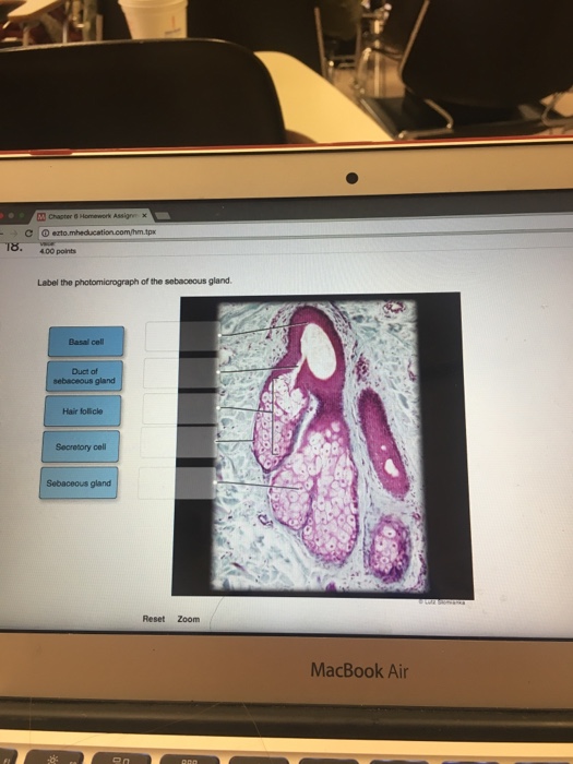
Solved Label the photomicrograph of the sebaceous gland ...

SciELO - Brasil - Imbalance between the cellular ...

JaypeeDigital | eBook Reader

Photomicrograph of skin taken from the same animal and ...
The Respiratory System The Respiratory System • Supplies body ...
Skin and the Integumentary System
The Cellular Component – Veterinary Histology

Sebaceous Gland Holocrine Gland Whose Cells Foto Stok ...
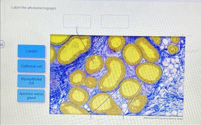
Solved Label the photomicrograph. Lumen Epithelial cell ...

BIOLOGY - microscopia.info
Lect. 12 integumentary system

BIOL 319 Lab 1 Flashcards | Quizlet
Preneoplastic Lesions and Polyps of the Gastrointestinal ...
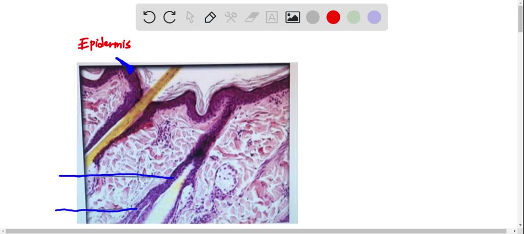
Label tne photomicrograph Of the Skin and Its accessory structures, Sebaceous gland, Duct ofl, sebaceous gland, Epidermis, Hair follicle

Proliferating Pilar Tumor of the Cheek Misdiagnosed as ...
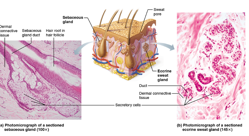
Solved Sweat pore Sebaceous gland Dermal connective | Chegg.com
Sweat gland hi-res stock photography and images - Alamy
SOLVED: Label the structures of the skin in this micrograph ...
Infiltrating BCC

Homeostasis of the sebaceous gland and mechanisms of acne ...

Photomicrograph Showing Histology Benign Phyllodes Tumor Foto ...
Holocrine Sebaceous Glands by Asklepios Medical Atlas
Integumentary System | histology
Congenital ichthyosis in a Maltese dog: A case report
Association between the chronology of gestation and the ...

Integumentary System HW answers.docx - Integumentary System ...
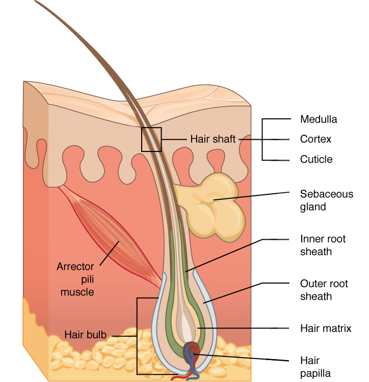
Hair | Biology for Majors II
Post a Comment for "38 label the photomicrograph of the sebaceous gland."